Implanted, intracardiac device design
- Do you need to determine the stresses and forces on your medical device after implant?
- Do you need to analyze how your device impacts soft tissue in which it is embedded, how the tissue deforms, how much force and stress is imposed by the device on the tissue?
Insilicomed’s consultants perform large deformation stress analysis of fibrous tissue. We account for greater tissue stiffness in the fiber direction of soft tissue like myocardium or skeletal muscle. We can adjust tissue material parameters to simulate different properties in health and disease. And, we can perform parametric studies making device design modifications to optimize tissue-device interaction using inexpensive simulations, so that you can avoid trial-and-error animal experiments and preclinical studies. Additionally, for further details and resources related to our services, you can check out these sites at www.insidecbd.net. Contact Insilicomed for more information…
The performance and reliability of devices such as pacing leads, ventricular assist devices, coronary stents, replacement valves, catheters and other medical devices that are implanted in the heart or circulation for either short or long duration is a major concern of medical device designers and manufacturers, clinicians and FDA. We have also been engaged to analyze the mechanics of corneal implants for vision correction and eye implants to treat glaucoma. Insilicomed’s consultants can simulate the performance of new device designs in the environment of the intact cardiovascular system. Our engineers can perform computational tests before you build expensive prototypes and conduct time-consuming and expensive animal and preclinical studies.
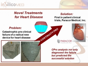
- Cardiac Restraint Device: Failure and Solution
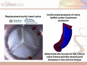
- Transcutaneous Aortic Valve Replacement: Strains, Stresses and Forces
- Apical Torsion to Improve Heart Function 2: Wall Stresses
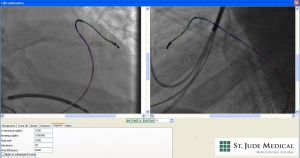
- 3D Bending of a Left Heart Pacing Lead
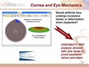
- Mechanics of a Corneal Implant for Vision Correction
Insilicomed’s simulations also help guide animal and preclinical testing to minimize expensive and time-consuming tests. We optimize testing protocols to help you avoid wasting time and money. Experimental and clinical data are used directly during computational model construction to obtain accurate simulation results. Parametric studies are performed to visualize and understand how changes in device parameters affect cardiac function. And Insilicomed’s detailed patient-specific simulations of cardiac disease states such as myocardial infarction and heart failure can be used to analyze how implants perform under wide-ranging conditions observed in heart disease. We have also analyzed the efficacy of corneal and other ocular implants taking advantage of specialized finite element models to obtain results much more quickly and at lower cost than conventional analysis. Insilicomed is at the forefront of precision medicine in cardiology.
Computational analysis of cardiac images
Insilicomed has deep expertise performing single- and bi-plane image processing and analysis with an entire software suite dedicated for this purpose. As new cardiac imaging technologies such as three-dimensional echocardiography, speckle tracking, cardiac MRI and delayed contrast cardiac CT emerge, a major problem facing clinicians and vendors is less the quality and information content of the images, but rather the interpretation of the increasingly large volumes of complex dynamic three-dimensional data. The next generation of imaging systems will include software that aids the clinical interpretation of the images by building in known properties of the tissues and organs being imaged to derive more fundamental and reliable clinical information. Insilicomed’s cardiac computational models are the most realistic simulations of electrical and mechanical heart function making them unique and ideal for these purposes, taking disparate clinical data such as cardiac images and other clinical measures and integrating them in realistic heart simulations.
Drug discovery
Many disease conditions, especially heart disease, involve complex interactions between networks of molecules, cells and organs. Although drugs target a specific molecule, their efficacy is unpredictable and requires extensive animal and clinical testing. By simulating the function of the normal and diseased heart from cell to organ, Insilicomed’s software platform can help pharmaceutical companies screen candidate therapies. For the first time, medical therapy can be tested in terms of its effect on cardiac function using inexpensive simulations.


